Japanese Pieris Grayanotoxins, Sodium-Channel Dysfunction, Severe Arrhythmias, Hypotension, and Respiratory Risk
Is Pieris Poisonous to Dogs, Cats, Horses, and Livestock?
Yes—Pieris, Pieris japonica (Thunb.) D.Don ex G.Don, is highly poisonous and potentially fatal to dogs, cats, horses, cattle, sheep, goats, pigs, llamas, alpacas, rabbits, guinea pigs, tortoises, birds, and other animals. Young and mature leaves, shoots, stems, bark, roots, sap, flower buds, open flowers, pollen, nectar, developing capsules, seeds, fresh clippings, wilted material, dried debris, and plant-contaminated feed should all be treated as toxic. Evergreen foliage and flower-bearing shoots create the greatest practical exposure because they remain abundant, visible, and accessible through much of the year.
Pieris contains grayanotoxins, a group of polyhydroxylated diterpenes that disrupt voltage-gated sodium channels in nerves, skeletal muscle, cardiac muscle, and autonomic tissue. The toxins prevent normal sodium-channel inactivation and hold excitable cells in an abnormally depolarized state. Poisoning can therefore combine excessive salivation, retching, vomiting, regurgitation, diarrhea, abdominal pain, bloat, tremors, and abnormal stimulation with weakness, conduction failure, paralysis, hypotension, altered consciousness, and respiratory collapse.
Cardiovascular abnormalities are central to severe poisoning. Sinus bradycardia and dangerously low blood pressure are characteristic, but tachycardia, junctional rhythms, atrioventricular block, ventricular ectopy, and other arrhythmias can also occur, and the rhythm may change during the illness. Weak pulses, pale mucous membranes, cold extremities, delayed capillary refill, reduced urine production, fainting, collapse, or reduced responsiveness indicates inadequate circulation even when the owner cannot determine the heart rate accurately.
Vomiting, regurgitation, impaired swallowing, recumbency, seizures, and ruminant bloat create major secondary hazards. Saliva, ruminal contents, food, drench material, or activated charcoal can enter the lungs when airway reflexes are impaired, and aspiration pneumonia may develop after the direct cardiovascular and neurologic signs begin improving. Published cases include confirmed grayanotoxin I poisoning in pet pigs, fatal and nonfatal goat outbreaks, poisoning in an African spurred tortoise, and a rapidly fatal alpaca case with severe pulmonary congestion and edema.
About this guide: This page provides general pet-poisoning information and cannot diagnose or treat an individual animal. For any suspected exposure, contact a veterinarian or animal poison-control service immediately. Do not induce vomiting, give medication, or attempt home decontamination unless directed by a veterinary professional.
Pieris
Pieris japonica (Thunb.) D.Don ex G.Don
- Andromeda japonica Thunb. — accepted homotypic botanical synonym and the source of the enduring common names Japanese Andromeda and Andromeda Japonica
- Pieris japonica var. japonica — accepted autonymous variety
- Pieris japonica var. yakushimensis T.Yamaz. — accepted variety native to Yakushima, Japan
Ericaceae — Heath or Heather Family
Pieris; Japanese Pieris; Japanese Andromeda; Andromeda Japonica; Lily-of-the-Valley Bush; Lily-of-the-Valley Shrub; Lily of the Valley Bush; Lily of the Valley Shrub; Japanese Fetterbush; Fetterbush; Asebi; Ashibi
Historical and taxonomic search variations include Andromeda japonica Thunb., Pieris japonica var. japonica, and Pieris japonica var. yakushimensis T.Yamaz.
Fetterbush is an ambiguous common name applied to Japanese Pieris, the separate native species Pieris floribunda, and toxic shrubs in the genera Lyonia and Leucothoe. Lily-of-the-Valley Bush is not true Lily of the Valley, Convallaria majalis; both are poisonous, but Pieris contains grayanotoxins while Convallaria contains cardiac glycosides. Japanese Pieris is identified by its dense evergreen woody habit, alternate glossy serrated leaves, bronze-to-red emerging foliage, pendent clusters of small urn-shaped white or pink flowers, dry five-part capsules, and the complete scientific label.
Grayanotoxins and Exact-Species Evidence
Japanese Pieris contains grayanotoxins, a family of highly oxygenated polycyclic diterpenes produced by several genera within Ericaceae. More than twenty-five related grayanotoxin structures have been described across heath-family plants, and individual species can contain several forms with different potency and tissue distribution. Grayanotoxin I and grayanotoxin III are among the principal clinically important isoforms, while grayanotoxin II is generally considered less potent but can occur with more active compounds.
Exact veterinary confirmation exists for grayanotoxin I in Pieris japonica. In two poisoned miniature pigs, investigators identified grayanotoxin I in the consumed shrub and in gastric contents, blood, liver, bile, kidneys, urine, lungs, and skeletal muscle through liquid chromatography–tandem mass spectrometry. This case confirms systemic absorption and distribution after whole-plant ingestion rather than merely inferring the toxin from family membership.
Historical toxin names can make the chemistry appear more complicated than it is. Grayanotoxin I has also been called andromedotoxin, acetylandromedol, rhodotoxin, and asebotoxin, while grayanotoxin III has been called andromedol and grayanotoxin II has been called andromedenol. These historical names should not be listed as though each were a separate toxic principle.
Modern phytochemical work on the genus Pieris has identified many structurally diverse grayanoids beyond the three traditional numbered toxins. Some possess analgesic, antifeedant, insecticidal, or other experimental activity, but their contribution to naturally occurring animal poisoning has not been separated compound by compound. Grayanotoxin I remains the strongest exact-species veterinary analytical anchor for Japanese Pieris.
Voltage-Gated Sodium Channels and Persistent Depolarization
Nerves and muscles depend on precisely timed opening, inactivation, and resetting of voltage-gated sodium channels. A channel normally opens briefly when an electrical impulse begins, permits sodium entry, then inactivates so the membrane can repolarize and respond to the next signal. Grayanotoxins bind preferentially to activated sodium channels and interfere with that normal inactivation process.
Binding stabilizes an abnormally open or activatable state, shifts channel activation toward more negative membrane potentials, and permits prolonged sodium entry. The cell remains depolarized and may initially discharge repetitively, producing salivation, retching, muscle twitching, tremors, abnormal autonomic activity, or tachycardia. Continued depolarization then prevents normal resetting and conduction, contributing to weakness, bradycardia, heart block, paralysis, reduced responsiveness, and respiratory failure.
Experimental sodium-channel research localizes important grayanotoxin interactions to transmembrane regions of the channel, including the D4S6 segment, and supports intracellular access to the binding region after channel activation. This molecular evidence explains the general mechanism but does not provide a safe leaf count for an animal. Natural poisoning involves several toxin isoforms, variable absorption, and species-specific physiology rather than a controlled laboratory concentration.
“Grayanotoxins work by binding to sodium channels in cell membranes. The binding unit is the group II receptor site, localized on a region of the sodium channel that is involved in the voltage-dependent activation and inactivation. These compounds prevent inactivation; thus, excitable cells (nerve and muscle) are maintained in a state of depolarization, during which entry of calcium into the cells may be facilitated. This action is similar to that exerted by the alkaloids of veratrum and aconite. All of the observed responses of skeletal and heart muscles, nerves, and the central nervous system are related to the membrane effects.”
United States Food and Drug Administration, Center for Food Safety and Applied Nutrition, Foodborne Pathogenic Microorganisms and Natural Toxins, 1992.
Autonomic, Cardiac, and Vascular Toxicity
Cardiac muscle, the sinoatrial node, the atrioventricular conduction system, autonomic nerves, and vascular smooth muscle are all vulnerable to abnormal sodium-channel behavior. Sinus bradycardia and hypotension are classic findings, but tachycardia, junctional rhythms, nodal rhythms, varying degrees of atrioventricular block, ventricular ectopy, and other rhythm disturbances can occur. The rate and rhythm can change as absorption, autonomic tone, dehydration, circulation, treatment, and toxin elimination evolve.
Grayanotoxin-related bradycardia is commonly associated with increased vagal activity and impaired pacemaker function. Hypotension may result from bradycardia, reduced cardiac output, abnormal vascular tone, vomiting or diarrhea, dehydration, and poor perfusion acting together. A normal or rapid heart rate does not prove that circulation is adequate because tachycardia can accompany shock.
Experimental work in feline cardiac Purkinje fibers showed that grayanotoxin III can alter cardiac electrical behavior and generate abnormal afterpotentials. The experiment helps explain ectopy and conduction instability but should not be converted into a prediction that every animal will develop one specific arrhythmia. Continuous ECG and blood-pressure monitoring are necessary because treatment depends on the rhythm and perfusion actually present.
Weak pulses, delayed capillary refill, pale mucous membranes, cold extremities, reduced urine production, apparent blindness, profound weakness, collapse, or altered awareness may reflect inadequate blood flow to the brain and other organs. Severe hypotension can become life-threatening even when gastrointestinal signs appear to be improving. Circulatory assessment must include heart rhythm, pulse quality, blood pressure, mucous-membrane color, mentation, temperature, and urine output together.
Gastrointestinal, Neuromuscular, and Respiratory Effects
Grayanotoxin disturbance of autonomic and excitable tissue commonly produces hypersalivation, lip smacking, tearing, retching, vomiting, regurgitation, abdominal pain, diarrhea, reduced appetite, abnormal gastrointestinal motility, and depression. Ruminants may develop repeated regurgitation, frothy salivation, ruminal atony, or bloat. Horses cannot vomit and may instead show salivation, repeated swallowing, colic, diarrhea, sweating, weakness, or abnormal behavior.
Skeletal-muscle and nervous-system effects can include twitching, tremors, stiffness, a wide-based stance, ataxia, inability to rise, paddling, opisthotonus, paralysis, seizures, stupor, or coma. The same sodium-channel disturbance can produce initial abnormal stimulation followed by inability to conduct or contract normally. Severe hypotension, electrolyte disturbance, hypoxia, and aspiration may intensify the neurologic picture.
Respiratory compromise can result from central depression, skeletal-muscle weakness, paralysis, poor circulation, pulmonary edema, severe abdominal distension, aspiration, or several mechanisms simultaneously. The fatal alpaca case included severe pulmonary congestion and edema together with white foamy material in the trachea and bronchi. In goat outbreaks, aspiration pneumonia remained a major cause of death after the direct grayanotoxin syndrome had begun improving.
Forced oral treatment increases danger when the animal is salivating, retching, weak, recumbent, neurologically abnormal, or unable to swallow normally. Activated charcoal, drench material, food, water, saliva, vomit, and ruminal contents can all enter the lungs. Airway protection and effective breathing take priority over decontamination once neurologic or respiratory signs develop.
All Plant Parts, Nectar, Honey, and Processed Material
Young and mature leaves, shoots, bark, stems, roots, sap, flower buds, open flowers, pollen, nectar, developing capsules, seeds, fresh clippings, wilted material, and dried ornamental debris should all be treated as poisonous. Exact toxin concentrations vary among tissues, seasons, cultivars, and individual shrubs, but no part has been established as safe animal food. Leaves and flowering shoots create the most frequent practical exposure because they are abundant and remain available on an evergreen shrub.
Drying, frost injury, pruning, and ordinary storage do not establish that the diterpene toxins have been destroyed. Clippings mixed into hay, bedding, silage, browse piles, municipal landscape waste, or compost can remain hazardous. Processing may make fragments more difficult to recognize and prevent animals from selecting around the plant.
Nectar from grayanotoxin-producing Ericaceae can be collected by bees, and honey dominated by toxic nectar can produce mad-honey poisoning. Most well-documented outbreaks involve regional Rhododendron species rather than Japanese Pieris acting alone, so the page should not claim that every Pieris flower produces dangerous honey. Honey, comb, nectar preparations, and homemade infusions from an uncertain Ericaceae source should nevertheless never be used as animal remedies.
Arbutin and other phenolic glycosides have been reported from some heath-family plants and may contribute to broader phytochemistry. They do not explain the rapid gastrointestinal, cardiovascular, neurologic, and respiratory syndrome documented after Pieris ingestion. Grayanotoxins remain the principal veterinary toxicants.
Dose Uncertainty and Species Variation
No dependable safe number of leaves, flowers, bites, or grams exists for dogs, cats, horses, livestock, pigs, tortoises, birds, or other animals. Experimental feeding of fresh Japanese Pieris equal to approximately 0.1% of one healthy goat’s body weight produced colic and nausea, while earlier natural goat cases included both fatal and nonfatal outcomes. That observation demonstrates potency in goats but is not a safe threshold, universal lethal dose, or calculation that should be transferred across species.
Plant toxicity varies with the grayanotoxin profile, leaf age, flower and shoot development, season, cultivar, environment, and individual shrub. Animal risk also depends on body size, species, age, health, stomach or rumen contents, amount consumed, repeated access, and the speed of treatment. Two animals sharing one exposure may develop different signs and outcomes.
The former practice of converting a goat percentage into an estimated amount for a dog should be avoided. It can falsely reassure an owner after a smaller ingestion and assumes equivalent plant chemistry, absorption, metabolism, and susceptibility between unrelated species. Every credible ingestion deserves case-specific veterinary or poison-control assessment.
Onset and Early Gastrointestinal Signs
Clinical signs usually begin within approximately one to four hours after ingestion, although delayed onset has been reported and depends on the amount, plant part, digestive state, and species. Early findings commonly include repeated lip movements, hypersalivation, foam around the mouth or snout, tearing, nasal discharge, apparent oral discomfort, gagging, retching, vomiting, regurgitation, abdominal pain, diarrhea, anorexia, and depression. These gastrointestinal signs may precede obvious cardiovascular or neurologic deterioration.
Horses cannot vomit, while cattle, sheep, goats, llamas, alpacas, and other ruminants may retch or regurgitate in a manner unusual enough to provide an important poisoning clue. Repeated swallowing, coughing, or feed and fluid returning through the mouth or nostrils may also indicate impaired swallowing or aspiration risk. An animal that continues eating or standing during the early phase is not necessarily stable.
Vomiting, regurgitation, and diarrhea can intensify the direct toxin effects through dehydration, electrolyte loss, aspiration, and reduced circulating volume. Ruminant gastrointestinal dysfunction can cause ruminal atony, accumulation of gas, and progressive abdominal distension. Severe bloat can restrict ventilation and venous return while increasing regurgitation risk.
Hypotension, Bradycardia, Tachycardia, and Arrhythmias
Blood pressure may fall as cardiac output, vascular tone, hydration, and autonomic regulation become impaired. Affected animals may appear weak, dizzy, reluctant to stand, cold through the ears or extremities, unusually quiet, apparently blind, or poorly responsive before collapse. Pale mucous membranes, delayed capillary refill, weak pulses, reduced urination, and loss of normal awareness indicate inadequate perfusion.
Sinus bradycardia is a characteristic finding, but it is not the only possible rhythm abnormality. Tachycardia, nodal or junctional rhythms, atrioventricular block, ventricular ectopy, and other arrhythmias can occur, and the rhythm may change during the course of poisoning. Owners cannot determine treatment from pulse rate alone because a rapid heart rate may reflect shock rather than adequate circulation.
Fainting, collapse, cold extremities, weak or irregular pulses, profound depression, or an abnormally slow or rapid heartbeat requires immediate ECG and blood-pressure assessment. The heart rate must be interpreted with perfusion, mucous-membrane color, respiratory status, temperature, and urine output. Temporary improvement in vomiting does not prove that conduction or blood pressure has normalized.
Neurologic and Neuromuscular Progression
Neurologic signs may begin with lethargy, unusual agitation, facial or muscle twitching, tremors, weakness, apparent visual impairment, a wide-based stance, or loss of coordination. The animal may stumble, sway, fall, become unable to rise, or progress to generalized recumbency. Poor cerebral perfusion and hypoxia can worsen the direct sodium-channel effects.
Severe poisoning may produce paddling, opisthotonus, limb paralysis, seizures, stupor, coma, or complete loss of voluntary muscle control. Animals may alternate between abnormal stimulation and profound weakness as excitable tissues remain depolarized and then fail to conduct normally. A quiet recumbent animal may therefore be deteriorating rather than resting.
People with grayanotoxin poisoning sometimes describe tingling or numbness around the mouth and extremities, but animals cannot report paresthesia. Face rubbing, pawing, unexplained agitation, abnormal limb responses, or apparent discomfort must be interpreted cautiously because nausea, weakness, pain, hypoperfusion, and neurologic dysfunction can produce similar behavior. Treatment should be guided by examination rather than assigning a sensation the animal cannot describe.
Respiratory Failure, Aspiration, and Pulmonary Complications
Respiratory abnormalities may result from central nervous system depression, skeletal-muscle weakness, paralysis, shock, pulmonary edema, severe abdominal distension, aspiration, or a combination of mechanisms. Tachypnea, shallow breathing, respiratory noise, labored effort, open-mouth breathing, irregular respirations, or blue-gray mucous membranes indicates an emergency. Weak ventilation can occur even when the animal is no longer tremoring.
Vomit, regurgitated feed, saliva, water, charcoal, and drench material can enter the lungs when airway protection is impaired. Coughing during or after vomiting, nasal discharge, fever, abnormal lung sounds, increasing respiratory effort, or renewed depression may indicate aspiration pneumonia. This complication can appear after the original cardiovascular and neurologic signs have begun improving.
Pulmonary congestion and edema were prominent in the fatal alpaca case. White foamy fluid filled the tracheal and bronchial lumens, and the lungs were dark red, congested, and edematous. Those findings demonstrate that fatal respiratory disease may become part of the syndrome rather than merely a consequence of visible vomiting.
Dogs, Cats, Horses, and Livestock
Dogs and cats may develop hypersalivation, repeated swallowing, vomiting, diarrhea, abdominal discomfort, depression, weakness, cold extremities, an abnormal heart rate, incoordination, tremors, seizures, or collapse. Exact companion-animal Pieris case literature is limited compared with goat and livestock evidence, so severity should be assessed from the actual exposure and clinical condition. A small animal can receive a large practical dose from only part of a branch.
Horses may show salivation, repeated swallowing, colic, diarrhea, sweating, weakness, trembling, ataxia, abnormal pulse quality, recumbency, convulsions, and death. They generally encounter the shrub through hedge trimmings, branches extending over a fence, nursery waste, storm debris, or mixed ornamental clippings. Because horses cannot vomit, absence of emesis does not indicate mild poisoning.
Goats, sheep, cattle, llamas, alpacas, and other ruminants may develop frothy salivation, retching, regurgitation, ruminal atony, bloat, apparent abdominal pain, anorexia, depression, tremors, staggering, paralysis, recumbency, and inability to stand. Several animals sharing one source may become ill at different times because intake differs. One symptomatic animal should trigger immediate removal and evaluation of the entire group.
Pigs may show smacking movements, foam around the snout, pale mucous membranes, tachycardia, tachypnea, abdominal tenderness, tremors, ataxia, and sudden lateral recumbency. In the confirmed pet-pig report, one animal recovered after supportive treatment while the other died during transport. The dead pig had few specific pathologic findings beyond consumed plant material, emphasizing the importance of toxicologic analysis and exposure history.
Rabbits, Guinea Pigs, Birds, and Reptiles
Rabbits and guinea pigs should never receive Pieris as browse, forage, bedding, or enrichment. They may develop salivation, food refusal, abdominal pain, diarrhea, reduced fecal production, weakness, tremors, loss of balance, recumbency, or abnormal breathing. These animals cannot vomit, so deterioration may be expressed through appetite, fecal output, posture, and neurologic function.
Companion birds and poultry may shred evergreen leaves, flowers, capsules, or clippings and can consume small pieces repeatedly. Regurgitation, diarrhea, weakness, poor balance, inability to perch, tremors, seizures, or respiratory changes requires avian veterinary care. Exact avian dose-response information for Japanese Pieris remains limited.
Direct reptile evidence exists. An African spurred tortoise developed severe abdominal pain, tenesmus, phallus prolapse, vocalization, hypersalivation, and distress after eating Japanese Pieris and required intensive veterinary treatment. The case demonstrates that reptiles cannot be assumed resistant merely because many toxicity lists were developed from mammalian data.
Duration, Recovery, and Signs of a Complicated Course
Limited exposures may resolve within several hours, and many successfully treated animals recover substantially within approximately one day. Larger doses can produce signs lasting several days, particularly when hypotension, arrhythmias, aspiration pneumonia, bloat, pulmonary edema, prolonged recumbency, or neurologic impairment develops. Elimination of the toxin does not immediately reverse every secondary complication.
Improvement should include normalizing heart rhythm, blood pressure, pulse quality, mucous-membrane color, hydration, urine production, breathing, strength, gait, appetite, and gastrointestinal function. The end of vomiting or tremors alone does not establish recovery. Continued weakness, coughing, fever, abdominal enlargement, abnormal pulse, or reduced responsiveness requires reassessment.
Cardiovascular collapse, respiratory failure, severe aspiration, uncontrolled seizures, coma, and death remain possible after major exposure. Prognosis improves with rapid recognition, airway protection, circulatory support, ECG-guided treatment, and prevention of aspiration. Delayed transport and forced oral remedies can worsen outcome.
Exact Botanical Identity, Classification, and Native Range
Japanese Pieris is Pieris japonica (Thunb.) D.Don ex G.Don, a woody broadleaf evergreen shrub or small tree in Ericaceae, the Heath or Heather Family. Carl Peter Thunberg originally described the species as Andromeda japonica, and George Don published the accepted combination in Pieris in 1834. The historical generic placement explains the persistent common names Japanese Andromeda and Andromeda Japonica.
Current botanical treatment recognizes one species-level synonym, Andromeda japonica, and two accepted varieties: var. japonica and var. yakushimensis. The native range includes southeastern and south-central China, central and southern Japan, and Taiwan. It grows primarily in temperate mountain thickets, woodland margins, and related acidic-soil habitats.
Widespread horticultural use has moved the shrub far beyond its native range. It is now common in foundation beds, woodland gardens, public landscapes, cemeteries, parks, schools, apartment complexes, commercial entrances, patios, and animal-facility landscaping. Toxic exposure can therefore occur in regions where Pieris would never grow naturally.
Growth Form, Leaves, Flowers, and Capsules
Typical landscape plants form dense upright or rounded shrubs approximately 2.5–3.5 meters tall, although dwarf cultivars remain much smaller and old specimens can become larger. Branches are woody and may create several levels of low accessible foliage. Dwarf forms and container-grown plants can place nearly the entire shrub at the muzzle height of dogs, miniature pigs, rabbits, goats, and young livestock.
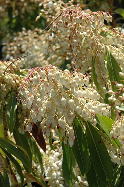
Leaves are alternate, simple, leathery, glossy, and dark green with finely serrated margins. New foliage often emerges bronze, copper, crimson, burgundy, pink, or bright red before maturing to green. The colorful tender shoots can be conspicuous and accessible when little other fresh vegetation is present.
Flower buds form in hanging clusters and can remain visible through winter before opening in late winter or spring. The small white, cream, or pink flowers are urn-shaped and arranged in pendent racemes resembling true Lily of the Valley. After flowering, dry capsules develop and may remain on the shrub while releasing small seeds.
Leaves, shoots, flowers, nectar, capsules, seeds, stems, bark, roots, sap, and discarded material all require control. A shrub does not become safe outside flowering season because evergreen leaves remain available. Seasonal cleanup can expose animals to larger quantities than they would reach on the intact plant.
Cultivars, Nursery Names, and Toxicity
Japanese Pieris is sold in many cultivars selected for size, leaf color, flower color, variegation, and flowering period. Common nursery names include ‘Mountain Fire’, ‘Mountain Snow’, ‘Forest Flame’, ‘Cavatine’, ‘Prelude’, ‘Katsura’, ‘Temple Bells’, ‘Dorothy Wyckoff’, ‘Flaming Silver’, ‘Valley Rose’, ‘Valley Valentine’, ‘Purity’, ‘Debutante’, and ‘Christmas Cheer’. Trade names may be displayed more prominently than the accepted species name.
No cultivar has been demonstrated to lack the relevant grayanotoxin hazard. Dwarf stature, pink flowers, variegated leaves, or red emerging foliage does not establish reduced toxicity. Every cultivar identified as Pieris japonica should be managed as poisonous.
‘Forest Flame’ and other commercially important plants may involve selection or hybrid ancestry within cultivated Pieris material. When the exact label is uncertain, the complete shrub should be preserved for identification. Similar management is appropriate while any Pieris or related grayanotoxin-containing Ericaceae remains possible.
Andromeda, Fetterbush, and Lily-of-the-Valley Confusion
Japanese Andromeda is an older common name derived from Andromeda japonica. True modern Andromeda species are botanically separate heath-family plants. Historical nursery records and landscape plans may still use Andromeda without making that distinction.
Fetterbush is applied to several toxic shrubs. It may identify Japanese Pieris, Mountain Pieris or Mountain Andromeda, Pieris floribunda, or species within Lyonia and Leucothoe. Several of those shrubs also contain grayanotoxins, so uncertainty does not make the exposure safe, but exact page attribution should remain botanically correct.
Lily-of-the-Valley Bush is not true Lily of the Valley, Convallaria majalis. Pieris is an evergreen woody shrub with grayanotoxins, while Convallaria is a herbaceous perennial containing cardiac glycosides. Both can produce vomiting, bradycardia, arrhythmias, weakness, and collapse, but detailed toxicologic assessment and treatment considerations differ.
Household, Landscape, Nursery, and Waste Exposure
Japanese Pieris is frequently planted beside foundations, porches, walkways, patios, fences, driveways, entrances, kennels, schools, veterinary clinics, public gardens, and apartment buildings. Low branches and hanging flower clusters can remain within easy reach. Container-grown shrubs may be pulled over by a dog or climbed by a cat.
Large-animal poisoning often follows landscaping activity rather than ordinary pasture grazing. Clippings may be thrown over a fence, fed deliberately as unfamiliar green browse, mixed into hay or bedding, or placed temporarily where escaped animals can reach them. Storm-damaged branches, transplanted shrubs, discarded nursery stock, cemetery arrangements, and seasonal cleanup create similar exposures.
Greenhouse and nursery waste can contain several Pieris cultivars, azaleas, rhododendrons, laurels, fertilizer, pesticide, wire, tags, plastic pots, and other hazardous material. A mixed load should be treated as an unknown toxic exposure until every component is identified. Apparently harmless mulch or compost may conceal leaves and flowers beneath the surface.
The plant’s bitterness does not provide dependable protection. Goats investigate woody browse, miniature pigs root through vegetation, puppies chew branches, tortoises consume unfamiliar ornamentals, and animals introduced to a new enclosure may sample plants despite having ordinary food available. Forage scarcity increases risk but is not required.
How Grayanotoxin Poisoning Can Change During One Case
Persistent sodium-channel activation does not mean that every tissue simply remains continuously stimulated. Initial abnormal depolarization may produce salivation, vomiting, regurgitation, twitching, tremors, tachycardia, or agitation. As cells become unable to reset and conduction fails, weakness, bradycardia, heart block, paralysis, reduced consciousness, and respiratory failure can follow.
Cardiovascular findings can change as autonomic tone, absorption, dehydration, circulation, and treatment evolve. One ECG snapshot cannot describe the entire illness. Continuous or repeated monitoring is appropriate when pulse rate, perfusion, mentation, or blood pressure is abnormal.
Secondary problems can continue after the direct toxin effect begins resolving. Aspiration pneumonia, pulmonary edema, bloat, recumbency injuries, electrolyte abnormalities, and kidney stress may determine the later outcome. Discharge decisions should therefore follow complete physiologic recovery rather than one temporarily normal heart rate.
Mad Honey and Xenophon’s Account
Grayanotoxin poisoning has been recognized for more than two thousand years. The best-known ancient account appears in Xenophon’s Anabasis, which describes Greek soldiers eating honey during the retreat of the Ten Thousand in the Black Sea region around 401–400 BCE. The honey is generally associated with nectar from toxic regional Rhododendron species rather than Japanese Pieris specifically, but the toxic mechanism is closely related.
“The number of bee hives was extraordinary, and all of the soldiers that ate of the honey combs lost their senses, vomited and were affected with purging, and none of them was able to stand upright; such as had eaten only a little were like men greatly intoxicated, and such as had eaten much were like mad men and some like persons at the point of death. They lay upon the ground, in consequence, in great numbers, as if there had been a defeat; and there was general dejection. The next day, no one of them was found dead; and they recovered their senses about the same hour they had lost them on the preceding day.”
Xenophon, Anabasis, account of the retreat of the Ten Thousand.
The account illustrates dose-dependent vomiting, diarrhea, altered awareness, inability to stand, and near-fatal collapse followed by recovery as exposure ended. It does not establish that every grayanotoxin exposure resolves without treatment. Animals consuming foliage can additionally develop aspiration, ruminal bloat, pulmonary complications, and fatal cardiovascular or respiratory failure.
Published Goat Evidence
A 1978 report described one fatal and one nonfatal natural goat case and then experimentally fed fresh Japanese Pieris equal to approximately 0.1% of a healthy goat’s body weight. Colic and nausea developed, and inhalation pneumonia was the principal necropsy finding in the fatal context. The study established that a relatively small amount can produce illness in a goat while also emphasizing aspiration as a major danger.
A 1979 report associated maternal Japanese Pieris poisoning with fetal mummification in a goat. One reproductive case does not establish pregnancy loss as the expected outcome of every ingestion. It does support strict prevention and veterinary monitoring when a pregnant animal is exposed.
A 1992 diagnostic report described Japanese Pieris toxicosis in a group of goats, reinforcing the syndrome of gastrointestinal distress and systemic illness after ornamental-shrub exposure. A 2001 report documented grayanotoxin poisoning in three goats and contributed important diagnostic and treatment evidence. Together, these reports establish exact-species goat risk across several decades.
Seven Goats, One Ram, and Intravenous Lipid Emulsion
In a later outbreak, seven goats and one ram escaped from a barn and consumed Japanese Pieris leaves. The animals developed regurgitation, obtundation, anorexia, apparent pain, and bloat. Initial supportive treatment included medication and activated charcoal; the ram and two goats recovered, while two goats died.
Three goats remained anorectic approximately thirty-six hours after exposure. One received intravenous lipid emulsion and improved within several hours, and the remaining two received the treatment the following day and appeared to improve. One of those animals later died from aspiration pneumonia.
The temporal improvement after lipid emulsion is clinically interesting because grayanotoxins are lipophilic, but the report did not establish a universal antidote, controlled comparison, or proven dose for every species. Intravenous lipid treatment can cause complications and must remain a veterinarian-directed adjunct. The delayed aspiration death also shows that visible neurologic or cardiovascular improvement does not end respiratory risk.
Confirmed Grayanotoxin I Poisoning in Two Pet Pigs
Two miniature pigs approximately two and a half years old and weighing about thirty and thirty-two kilograms developed sudden tremors and ataxia after access to Japanese Pieris. Both progressed to lateral recumbency and repeatedly smacked their mouths with foam around the snout. Pale oral mucous membranes, tachycardia, tachypnea, abdominal tenderness, and apparent pain were documented.
The female pig received supportive fluid treatment and recovered within several hours. The male received initial symptomatic care but died during transport. Postmortem examination found plant material within a severely congested section of small intestine but no single distinctive lesion that would have diagnosed the poisoning independently.
Investigators identified grayanotoxin I in the consumed plant, gastric contents, blood, liver, bile, kidneys, urine, lungs, and skeletal muscle through HPLC-MS/MS. This report is especially important because it chemically confirmed exact-species systemic poisoning. It also demonstrates that similar animals with apparently similar access can have very different outcomes.
Fatal Japanese Pieris Poisoning in an Alpaca
A ten-year-old female alpaca was introduced into a newly constructed outdoor enclosure and was found later the same day in lateral recumbency with profuse frothy salivation and inability to stand. Intravenous fluids, gastric intubation, attempted lavage, activated charcoal, oxygen, anticonvulsant treatment, and supportive care were provided. The condition continued deteriorating.
Vomiting, paddling movements, opisthotonus, and an abnormal Cheyne–Stokes respiratory pattern developed, and the alpaca died approximately four hours after clinical signs were first recognized. Necropsy found green gastric contents containing ten leaves identified morphologically as Japanese Pieris. Browsed shrubs were present inside the enclosure.
The lungs were severely congested and edematous, with white foamy fluid in the trachea and bronchi. Hemorrhages occurred around the stomach and duodenum, and marked congestion affected the liver, spleen, and kidneys. The report was diagnosed from the clinical course, recovered leaves, enclosure evidence, and pathology rather than direct grayanotoxin measurement and is therefore best described as a strongly supported suspected or presumptive exact-species poisoning.
African Spurred Tortoise Case
A four-year-old male African spurred tortoise developed severe abdominal pain after eating Japanese Pieris. Tenesmus with protrusion of the phallus, vocalization, hypersalivation, and marked distress were reported. Intensive reptile veterinary treatment was required.
The case is important because plant poisoning in reptiles is poorly documented and many reptile plant lists are extrapolated from mammals. It demonstrates that a tortoise can become clinically ill from Japanese Pieris and should not be presumed resistant. Tortoise yards and planted herbivore enclosures should not contain this shrub.
Dogs, Cats, Horses, and Other Animal Groups
Dogs may chew low branches, carry clipped stems, raid landscape-waste piles, or pull a container-grown shrub from a porch. Cats may bite young foliage or flowers and groom sap or fragments from the paws and coat. Exact dog-and-cat case literature is less extensive than goat evidence, but the sodium-channel mechanism and cross-species reports justify urgent assessment after credible ingestion.
Horses generally encounter Japanese Pieris through hedge trimmings, landscaping debris, branches growing across fences, discarded nursery plants, or mixed ornamental waste. They cannot vomit and may instead develop salivation, colic, diarrhea, sweating, weakness, tremors, abnormal pulse, ataxia, recumbency, seizures, and respiratory failure. Drenching an uncoordinated horse creates a severe aspiration hazard.
Cattle, sheep, goats, llamas, and alpacas may consume the plant when clippings are mixed with desirable browse or when animals escape into landscaped areas. Pigs may root through whole shrubs or discarded material. Rabbits, guinea pigs, birds, and tortoises can receive a substantial dose relative to body size and should never be offered the plant as forage or enrichment.
Diagnosis, Differential Diagnosis, and Prognosis
Diagnosis combines exact plant identification, credible access, estimated amount, onset, gastrointestinal signs, cardiovascular findings, neurologic progression, and respiratory status. Preserve whole branches bearing mature leaves, colorful shoots, flowers or capsules, together with labels, photographs, vomit, regurgitated material, stomach contents, feed, and clippings. A complete sample is more useful than one detached leaf.
Veterinary assessment may include ECG, blood pressure, pulse quality, mucous-membrane color, capillary refill, hydration, temperature, respiratory effort, abdominal distension, rumen activity, neurologic function, swallowing, oxygenation, blood gases, electrolytes, kidney values, muscle enzymes, and thoracic imaging. There is no routine clinic-side grayanotoxin test. Specialized laboratories can analyze plant material and biological samples, but emergency treatment cannot wait for those results.
Differential diagnoses include azalea, rhododendron, Mountain Laurel, Sheep Laurel, Lyonia, Leucothoe, aconite, Veratrum, yew, Oleander, true Lily of the Valley, organophosphate or carbamate pesticides, metaldehyde, nicotine, cardiac medication, cannabis, primary cardiac disease, severe electrolyte disorders, bloat, and aspiration. Common-name confusion is especially important because several unrelated plants may be called laurel, lily of the valley, fetterbush, or andromeda.
Small exposures limited to transient gastrointestinal signs generally have a favorable prognosis when cardiovascular and respiratory monitoring remains normal. Significant hypotension, heart block, serious arrhythmias, paralysis, seizures, bloat, pulmonary edema, aspiration, coma, or respiratory failure creates a guarded prognosis. Fatal outcomes are documented, but rapid aggressive supportive care can produce complete recovery.
Prevention
Japanese Pieris should not be planted where browsing animals, habitual plant-chewing pets, miniature pigs, rabbits, poultry, tortoises, or zoo animals can reach it. Inspect new enclosures before introducing animals and account for branches growing through or over fences. Dwarf plants and containers may be more accessible than mature foundation shrubs.
Place every clipping directly into a closed animal-inaccessible container. Never throw Pieris, azalea, rhododendron, laurel, fetterbush, or mixed ornamental debris into a pasture, livestock pen, poultry run, rabbit area, compost pile, or hay supply. Storm debris and uprooted shrubs require the same control.
Educate landscapers, groundskeepers, neighbors, nursery workers, animal caretakers, and visitors that fresh green ornamental branches are not safe browse. Retain plant labels and property records so a future exposure can be identified quickly. Removal remains the safest option where reliable separation cannot be maintained.
Immediate Owner Actions
Any known or suspected Japanese Pieris ingestion warrants prompt veterinary or animal poison-control guidance. Cardiovascular and neurologic abnormalities may follow an early period dominated by drooling, vomiting, regurgitation, or diarrhea. The animal should not remain at home merely because it still appears alert or can stand.
- Remove access immediately: Prevent contact with the shrub, leaves, flowers, nectar, capsules, seeds, roots, clippings, contaminated feed, compost, and gastrointestinal material.
- Preserve a complete specimen: Save a branch with mature leaves, young growth, flowers or capsules when available, together with photographs and the nursery label.
- Estimate the maximum exposure: Record the animal’s weight, earliest possible ingestion time, amount missing, plant parts involved, and whether several animals had access.
- Keep the animal quiet: Restrict exertion because hypotension, arrhythmia, weakness, and collapse can worsen during excitement.
- Contact professional help immediately: Do not wait for tremors, paralysis, respiratory distress, or loss of consciousness.
- Remove other animals: Separate all housemates, herd mates, flock mates, and poultry from the same source and from vomited or regurgitated material.
What Not to Do at Home
Decontamination may be useful after a recent exposure, but it must be selected according to species, elapsed time, symptoms, swallowing ability, airway protection, and cardiovascular stability. A procedure appropriate for a neurologically normal dog may be dangerous or impossible in a horse, ruminant, rabbit, tortoise, weak animal, or patient already vomiting or regurgitating. Home treatment should never delay transportation.
- Do not give hydrogen peroxide: It can cause prolonged vomiting, gastric and esophageal injury, dehydration, and aspiration and must never be used automatically.
- Do not force activated charcoal: A salivating, retching, vomiting, weak, recumbent, tremoring, seizing, bloated, or poorly swallowing animal can inhale charcoal.
- Do not drench horses or livestock: Grayanotoxin poisoning may impair swallowing and increase regurgitation and aspiration.
- Do not give atropine at home: A slow pulse does not prove atropine is appropriate, and some patients develop tachycardia or another rhythm disturbance.
- Do not give antiarrhythmic or heart medication: Treatment must follow an ECG-confirmed rhythm and measured perfusion.
- Do not give anti-diarrheal or human medication: These products do not neutralize grayanotoxins and may worsen gastrointestinal or cardiovascular management.
- Do not force food, milk, oil, or water: None is an antidote, and forced swallowing can produce aspiration.
- Do not give honey: Honey is not an antidote and an uncertain Ericaceae-derived product may itself contain grayanotoxins.
Emergency Signs and Safe Transport
- Gastrointestinal emergency: Repeated vomiting, retching, regurgitation, profuse diarrhea, severe abdominal pain, inability to retain water, or blood in gastrointestinal material.
- Cardiovascular emergency: An abnormally slow, rapid, weak, or irregular pulse; pale gums; cold extremities; fainting; profound weakness; or collapse.
- Neurologic emergency: Twitching, tremors, staggering, apparent blindness, inability to stand, paddling, opisthotonus, seizures, paralysis, stupor, or coma.
- Respiratory emergency: Rapid, shallow, labored, noisy, weak, or irregular breathing; blue-gray mucous membranes; coughing after vomiting; or foam from the mouth or nostrils.
- Ruminant bloat: Progressive left-sided abdominal enlargement, repeated lying down, kicking at the abdomen, respiratory distress, or inability to belch.
- Multiple exposed animals: Treat the whole group as potentially affected even when only one animal is visibly ill.
Call the receiving clinic before departure and report suspected Pieris japonica or grayanotoxin poisoning. Use a carrier, blanket, board, or stretcher rather than forcing a weak, hypotensive, ataxic, or partially paralyzed animal to walk. Position the head so saliva, vomit, or regurgitated material can drain from the mouth, and bring the plant, label, photographs, feed, and safely contained gastrointestinal samples.
Veterinary Stabilization and Decontamination
Airway, breathing, circulation, neurologic status, and dangerous bloat take priority over gastrointestinal decontamination. Continuous ECG and repeated blood-pressure assessment are central because the cardiac rhythm and perfusion can change during illness. Oxygen, suctioning, intubation, assisted ventilation, vascular access, and emergency rumen decompression may be needed before plant removal is attempted.
A veterinarian may induce vomiting after a recent meaningful ingestion in an alert, neurologically normal dog or cat with intact airway reflexes. Pieris leaves may remain within the stomach long enough for professionally controlled emesis to remain useful beyond the very narrow window associated with some rapidly absorbed poisons. Once marked weakness, tremors, seizures, altered consciousness, spontaneous vomiting, or impaired swallowing develops, emesis is unsafe.
Veterinarian-administered activated charcoal may reduce absorption when plant material remains in the gastrointestinal tract and the airway is protected. Gastric lavage may be considered after a serious recent exposure under anesthesia with endotracheal intubation. Charcoal, lavage, cathartics, and enemas are not harmless and must not delay treatment of hypotension, arrhythmias, bloat, respiratory distress, or seizures.
Cardiovascular, Neurologic, and Respiratory Treatment
There is no specific antidote that directly removes grayanotoxin from sodium channels. Treatment supports the animal while the toxin is metabolized and eliminated and while secondary complications are controlled. Drug selection depends on the measured rhythm, blood pressure, perfusion, respiratory function, and species.
- Support circulation: Intravenous fluids are adjusted to hydration, blood pressure, cardiac function, pulmonary findings, and urine production.
- Treat significant bradycardia: Atropine may be used when ECG findings, pulse quality, blood pressure, and perfusion demonstrate clinical need.
- Treat other arrhythmias: Lidocaine, procainamide, or another veterinarian-selected agent may be considered according to the documented rhythm.
- Control tremors and seizures: Methocarbamol, benzodiazepines, barbiturates, propofol, or other clinician-selected treatments may be required.
- Protect the airway: Oxygen, suctioning, intubation, ventilation, and aspiration treatment may be necessary.
- Manage bloat: Ruminants may require urgent decompression and restoration of rumen function.
- Correct secondary abnormalities: Electrolyte disturbance, acid-base imbalance, dehydration, hypothermia, hyperthermia, and reduced urine production are treated from measured findings.
Intravenous Lipid Emulsion and Treatment Limitations
Intravenous lipid emulsion was followed by rapid improvement in several goats that remained anorectic after ordinary supportive treatment. The proposed rationale is that a lipid phase may bind or redistribute lipophilic grayanotoxins. The case report did not include a controlled untreated comparison and therefore cannot prove that lipid emulsion alone caused recovery.
Intravenous lipid emulsion is not a universal antidote and should not be administered outside veterinary supervision. It can interfere with laboratory testing, alter blood viscosity, and contribute to metabolic, pancreatic, pulmonary, or other complications. Species experience and evidence remain limited.
The miniature-pig report demonstrates that apparently similar exposures can produce different outcomes: one pig recovered after fluid support, while the other died during transport. The alpaca case demonstrates that charcoal and attempted lavage cannot substitute for successful cardiovascular and respiratory stabilization. The goat outbreak demonstrates that aspiration pneumonia can cause delayed death after apparent improvement.
Species-Specific Precautions
Dogs and cats should be monitored beyond the end of vomiting for pulse quality, blood pressure, breathing, strength, gait, and aspiration. Cats with prolonged food refusal require nutritional planning, but force-feeding is unsafe during nausea, altered awareness, or impaired swallowing. Any renewed coughing, fever, weakness, or collapse requires reassessment.
Horses should not be drenched or forced to walk when weak or uncoordinated. Salivation, colic, diarrhea, sweating, abnormal pulse, trembling, ataxia, recumbency, or respiratory change requires immediate large-animal care. Branches and clippings should be removed from the entire paddock while other horses are observed.
Ruminants require monitoring for regurgitation, bloat, rumen contractions, respiratory effort, appetite, manure production, and aspiration. Affected animals should be positioned and handled to reduce inhalation of ruminal contents. Apparently normal animals sharing the source require continued observation because onset and intake differ.
Rabbits, guinea pigs, birds, and tortoises should not undergo home-induced vomiting. Food refusal, reduced feces, regurgitation, poor balance, inability to perch, tremors, abnormal breathing, or severe abdominal discomfort requires specialized veterinary care. Force-feeding or syringe fluids are unsafe until swallowing, gastrointestinal function, and respiratory status have been assessed.
Monitoring, Prognosis, and Prevention
Heart rate, rhythm, pulse quality, blood pressure, mucous-membrane color, capillary refill, breathing, hydration, urine production, strength, gait, gastrointestinal function, and rumen activity should normalize together. The end of salivation, vomiting, or tremors does not prove that atrioventricular block, hypotension, aspiration, pulmonary edema, or bloat has resolved. Observation should continue until the patient remains stable without rescue medication or intensive support.
- Monitor the heart: Rhythm and rate must remain compatible with normal blood pressure and tissue perfusion.
- Monitor breathing: Coughing, fever, nasal discharge, increased effort, or renewed depression may indicate aspiration pneumonia.
- Monitor hydration and urine: Drinking, gum moisture, urine production, and kidney measurements should recover as circulation improves.
- Monitor neurologic function: Strength, gait, vision, mentation, tremors, and ability to rise should improve steadily.
- Monitor ruminants for bloat: Abdominal size, rumen contractions, appetite, regurgitation, and manure production require continued assessment.
Small exposures limited to temporary gastrointestinal signs generally have a favorable prognosis. Significant hypotension, heart block, serious arrhythmias, paralysis, seizures, bloat, pulmonary edema, aspiration, coma, or respiratory failure creates a guarded prognosis, although aggressive treatment can still be successful. Remove Japanese Pieris from animal enclosures, inspect new paddocks and zoo exhibits before use, prevent branches from crossing fences, and place every clipping directly into a closed animal-inaccessible container.
Frequently Asked Questions About Pieris and Animal Poisoning
Is Japanese Pieris poisonous to dogs and cats?
Yes. Pieris japonica contains grayanotoxins that can cause drooling, vomiting, diarrhea, abdominal pain, depression, weakness, hypotension, bradycardia or tachycardia, arrhythmias, incoordination, tremors, seizures, paralysis, collapse, and death. Exact dog-and-cat case literature is less extensive than goat evidence, but the toxin mechanism and cross-species severity justify urgent assessment after credible ingestion. A small animal can receive a meaningful practical dose from only part of a branch.
Is Pieris poisonous to horses and livestock?
Yes. Horses, cattle, sheep, goats, pigs, llamas, alpacas, and other livestock can become severely or fatally poisoned. Exposure commonly follows access to hedge clippings, nursery waste, storm debris, escaped landscape shrubs, branches extending over fences, or plant material offered deliberately as browse. Exact cases include goats, sheep, miniature pigs, and an alpaca.
Which parts of Japanese Pieris are poisonous?
Young and mature leaves, shoots, stems, bark, roots, sap, flower buds, open flowers, pollen, nectar, developing capsules, seeds, fresh clippings, wilted material, and dried debris should all be treated as poisonous. Leaves and flower-bearing shoots create the greatest practical exposure because they are abundant and accessible. Drying, frost, and ordinary composting do not establish safety.
What is the accepted scientific name?
The accepted name is Pieris japonica (Thunb.) D.Don ex G.Don. Andromeda japonica Thunb. is its accepted botanical synonym and explains the common names Japanese Andromeda and Andromeda Japonica. Current botanical treatment also recognizes var. japonica and var. yakushimensis. Nursery labels may emphasize a cultivar name instead of the species.
Is Lily-of-the-Valley Bush the same as true Lily of the Valley?
No. Lily-of-the-Valley Bush is Japanese Pieris and contains grayanotoxins. True Lily of the Valley is Convallaria majalis and contains cardiac glycosides. Both can cause vomiting, bradycardia, arrhythmias, weakness, and collapse, but they are unrelated plants with different toxin chemistry. Preserve the complete plant rather than relying on the similar common name.
Is Fetterbush always Japanese Pieris?
No. Fetterbush may refer to Japanese Pieris, Pieris floribunda, or toxic shrubs in the genera Lyonia and Leucothoe. Several of these plants also contain grayanotoxins, so an uncertain identification still warrants urgent veterinary guidance. The complete branch, leaves, flowers, fruit, label, and growth form should be preserved.
What toxin is in Japanese Pieris?
The principal toxins are grayanotoxins, especially grayanotoxin I and grayanotoxin III. Grayanotoxin II is generally considered less potent. Grayanotoxin I has also been called andromedotoxin, acetylandromedol, rhodotoxin, and asebotoxin, but those are historical names for the same compound rather than separate toxins. Grayanotoxin I has been chemically confirmed in poisoned pet pigs.
How do grayanotoxins affect nerves and the heart?
They bind to activated voltage-gated sodium channels and interfere with normal channel inactivation. Nerve and muscle cells remain abnormally depolarized and cannot reset or conduct normally. The resulting disturbance affects the gastrointestinal tract, skeletal muscles, autonomic nerves, blood vessels, brain, cardiac conduction system, and heart muscle. Initial stimulation may be followed by weakness and conduction failure.
Can Pieris cause both a slow and a rapid heartbeat?
Yes. Sinus bradycardia and hypotension are characteristic, but tachycardia, junctional rhythms, atrioventricular block, ventricular ectopy, and other arrhythmias may occur. The rhythm can change during the illness. Treatment must be based on ECG, blood pressure, pulse quality, mucous-membrane color, and perfusion rather than the owner’s impression of pulse rate alone.
How quickly do signs begin?
Signs often begin within approximately one to four hours, although onset can be delayed. Early findings may consist only of drooling, lip movements, vomiting, regurgitation, diarrhea, abdominal pain, or depression. Cardiovascular and neurologic abnormalities can follow as absorption progresses. A normal appearance immediately after ingestion does not establish safety.
Can Pieris cause bloat in goats or cattle?
Yes. Abnormal gastrointestinal motility, ruminal atony, regurgitation, and recumbency may contribute to gas accumulation. Severe bloat can restrict breathing and circulation while increasing aspiration risk. Progressive left-sided abdominal enlargement, repeated lying down, respiratory distress, or inability to belch requires immediate large-animal veterinary treatment.
Can Pieris cause seizures, paralysis, or coma?
Yes. Larger exposures may produce twitching, tremors, ataxia, inability to stand, limb paralysis, paddling, opisthotonus, seizures, stupor, coma, and respiratory failure. Severe hypotension, electrolyte changes, and hypoxia can intensify the direct toxin effects. These findings require immediate emergency care and airway monitoring.
How much Pieris does it take to poison an animal?
No universal safe or lethal amount exists. Fresh foliage equal to approximately 0.1% of one goat’s body weight produced illness experimentally, but that figure is not a safety threshold and cannot be converted reliably to dogs, cats, horses, pigs, or other species. Plant chemistry and animal susceptibility vary. Every confirmed ingestion deserves case-specific professional assessment.
Have pet pigs been poisoned by Japanese Pieris?
Yes. Two miniature pigs developed pale mucous membranes, tachycardia, tachypnea, hypersalivation, tremors, ataxia, abdominal pain, and lateral recumbency. One recovered after supportive care, while the other died. Grayanotoxin I was identified in the plant, gastrointestinal contents, blood, liver, bile, kidneys, urine, lungs, and skeletal muscle.
Has Japanese Pieris poisoned a tortoise?
Yes. An African spurred tortoise developed severe abdominal pain, tenesmus, phallus protrusion, vocalization, hypersalivation, and distress after eating Japanese Pieris. The case required intensive reptile veterinary care and was the first published report of a Pieris species poisoning a reptile. Tortoise enclosures should not contain this shrub.
Has Japanese Pieris caused fatal alpaca poisoning?
Yes. A zoo-housed alpaca developed profuse frothy salivation, recumbency, vomiting, paddling, opisthotonus, abnormal respiration, and death within approximately four hours. Ten Pieris leaves were recovered from the stomach, and browsed shrubs were present inside the enclosure. Necropsy showed severe pulmonary congestion and edema with foamy fluid in the airways.
Should I make my dog or cat vomit?
Do not induce vomiting unless a veterinarian or animal poison-control professional specifically directs it after assessing the patient. Professional emesis may be appropriate after a recent exposure in an alert, neurologically normal dog or cat with intact airway reflexes. Spontaneous vomiting, weakness, tremors, altered awareness, or impaired swallowing creates a serious aspiration risk. Hydrogen peroxide should never be used automatically.
Should I give activated charcoal?
Do not force charcoal at home. A veterinarian may administer it when unabsorbed plant material remains and the airway is protected. A salivating, retching, vomiting, weak, bloated, recumbent, tremoring, seizing, or poorly swallowing animal can inhale charcoal and develop severe lung injury. Charcoal cannot correct an arrhythmia, hypotension, paralysis, or respiratory failure.
Is atropine an antidote?
No. Atropine may improve clinically important bradycardia when ECG, pulse quality, blood pressure, and perfusion show that it is appropriate. It does not remove or neutralize grayanotoxin. Some poisoned animals develop tachycardia or another arrhythmia, so unsupervised atropine can be inappropriate or dangerous.
Does intravenous lipid emulsion cure Pieris poisoning?
It is not a proven universal antidote. Several goats improved after veterinarian-administered intravenous lipid emulsion, but the report did not include a controlled comparison proving that lipid therapy alone caused recovery. One goat that initially improved later died from aspiration pneumonia. The treatment remains a veterinarian-selected adjunct with potential complications.
How do veterinarians treat Pieris poisoning?
Treatment may include controlled decontamination, intravenous fluids, continuous ECG and blood-pressure monitoring, atropine for appropriate bradycardia, rhythm-specific antiarrhythmic treatment, antiemetics, tremor or seizure control, oxygen, airway protection, ventilation, electrolyte correction, bloat relief, and treatment of aspiration pneumonia. There is no single antidote. Therapy follows the animal’s measured cardiovascular, neurologic, respiratory, and gastrointestinal abnormalities.
What is the prognosis?
Small exposures limited to temporary gastrointestinal signs generally have a favorable prognosis when blood pressure, rhythm, breathing, and neurologic function remain normal. The outlook becomes guarded with significant hypotension, heart block, serious arrhythmias, paralysis, seizures, bloat, pulmonary edema, aspiration, coma, or respiratory failure. Fatal cases are documented, but rapid aggressive supportive care can also produce complete recovery.